Thème d'affichage
Langue / Language
Join the FBKC
Create your free account and access all our services: pedigree requests, ratings, health tracking and more!
100% free & secure
A question about our services? Contact us directly!
Write on WhatsAppCreate your free account and access all our services: pedigree requests, ratings, health tracking and more!
100% free & secure
Dysplasia = Malformation or abnormality in the development of a tissue or organ resulting from a disorder of embryogenesis.
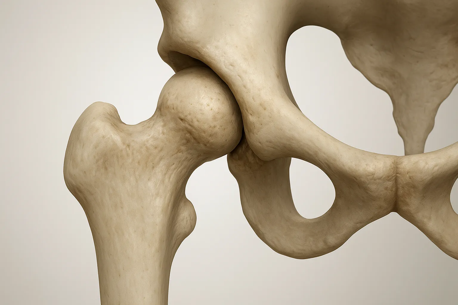
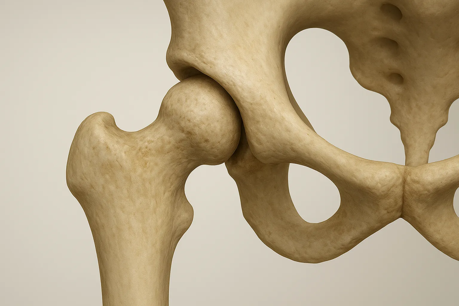
Hip (coxofemoral) dysplasia is therefore an abnormality of the hip joint resulting from a malformation or anomaly of one of the components of that joint.
- Articular surfaces: - the femoral head
- the acetabulum
- the coxofemoral ligament connecting the two articular surfaces
- the joint capsule containing synovial fluid
(not forgetting the muscles surrounding the joint)
Dysplasia is therefore an abnormality in the stability of the joint, initially due to excessive joint laxity (i.e. abnormality of the joint capsule and ligament, as well as muscular support), which leads to bone deformities, microfractures, and ultimately the development of arthritis. The phenomenon is self-aggravating.
In a normal, healthy hip, the articular surfaces fit together perfectly: they are geometrically complementary to one another.
It is the coxofemoral ligament and the joint capsule that keep one engaged within the other (refer to the diagram above: if the muscles and ligament are removed, the femur 'drops').
You can take a dysplasia radiograph on a puppy if you have concerns about the state of the hips, but for official grading, the dog must be at least 12 months old for the radiograph to be valid.
- A hip where the coxofemoral ligament is too 'long'. There is then excessive joint laxity. Even if the articular surfaces are perfectly matched to one another, the femur is not held in the acetabulum by the ligament, making the joint unstable again: the poorly supported femur will knock against the acetabulum with each step, creating microtrauma to the bone and therefore arthritis, and the hip subluxates or dislocates.
- Under the effect of these abnormal movements of the femur relative to the acetabulum, the bony surfaces become deformed: they no longer fit together because one (or both) surfaces are no longer geometrically matched to the other (for example, an acetabulum that is too flat, a femoral head that is not round enough... try fitting a square into a circle).
This leads to a problem of congruence, meaning that when the hip is in motion it is even less stable — the femur does not 'glide' perfectly within the acetabulum. This also creates friction of the femoral head within the acetabulum and therefore bone microtrauma, which in turn leads to arthritis.
Furthermore, the coverage of the femoral head by the acetabulum is inadequate: the acetabulum does not sufficiently 'surround' the femoral head, which 'allows' the hip to subluxate or dislocate — meaning the femoral head will partially or fully exit the acetabulum during certain movements. This again causes bone trauma and arthritis.
Of course, the muscles attempt to compensate for the hip's shortcomings: they do their best to compensate for the ligament defect by holding the femur in the acetabulum. They provide better joint stability by limiting subluxations. This explains why a very muscular dog will not necessarily show clinical signs of CHD.
Laxity is the cause of bone deformities and the formation of arthritis. In some dogs, this ligament laxity exists but is 'masked' by the action of the muscles, which correctly maintain the hip and prevent this laxity from being expressed, so that bone deformities do not appear. This is known as passive laxity. In other dogs with too much laxity, the muscles are not sufficient to stabilise the joint and bone deformities appear. This is known as active laxity.
Hip dysplasia is a disease that depends on a polygenic system and can be influenced by the external environment.
Genetics: This disease depends on quantitative genetics. In other words, a certain number of deleterious (abnormal) alleles are required for the disease to appear: it is a threshold disease. When this threshold is exceeded, if there are even more deleterious alleles, the symptoms worsen.
To keep things simple: a dog has two copies of each gene (as we do), these two copies being called the alleles of the gene. In a given population, there can be thousands of different alleles for the same gene — for example, the 'eye colour' gene has alleles 'blue', 'brown', 'black', 'red', 'green', etc.
The genetic determinism of CHD is therefore very complex: the deleterious alleles and even the genes involved are not known. The fact that many genes are involved complicates the problem: by looking at the results of the parents, we cannot say whether the offspring will be dysplastic or not.
The environment: on top of the genetic basis, environmental factors during growth are added: diet (an excess or deficiency of certain compounds), growth rate, the dog's weight, the dog's activity, etc. For example, a genetically healthy dog that grows at full speed while being 15 kilos overweight and covering 20 kilometres a day may become dysplastic.
Pain! The dog experiences pain during and/or after exertion: at rest, arthritis is painful. Of course, progression occurs over several months or even years. Disability partly due to pain: the more pain the dog is in, the less effort it places on its hip (less muscle use) and therefore the more pain it feels and the less flexible it becomes.
On clinical examination: the dog shows pain when the hip is manipulated. Extending the hip backwards, flexing it forwards or abducting the leg to the side all elicit pain.
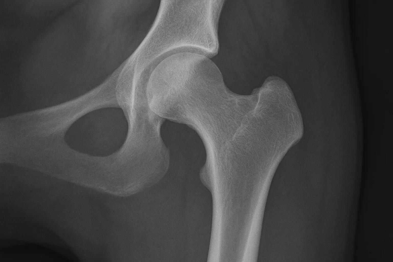
On radiography: a radiograph is taken with the hips extended backwards and the kneecaps at the zenith (pointing upwards): this position highlights the (sub)luxation of the femoral heads if there is a problem. When reading the radiograph, several things are assessed: the articular surfaces (regularity, congruence...), the joint space and the (sub)luxation of the hip.
Grade A: No sign of dysplasia, perfect congruence and coaptation, NORBERG-OLSSON angle > 105°
Grade B: Either the NORBERG-OLSSON angle is between 105° and 100° with perfect or near-normal coaptation and congruence, or the NORBERG-OLSSON angle is greater than 105° with more or less good congruence.
Grade C: Mild dysplasia. Angle between 100° and 105° and average congruence.
Grade D: Moderate dysplasia. Angle between 100° and 90° and congruence is truly poor. This stage is often accompanied by flattening of the acetabulum and the possibility of arthritis, though this is not systematic.
Grade E: Severe dysplasia. There is (sub)luxation of the femoral head and the angle is less than 90°. This stage is often accompanied, in addition to the signs of stage D, by an abnormality in the conformation of the femoral head.
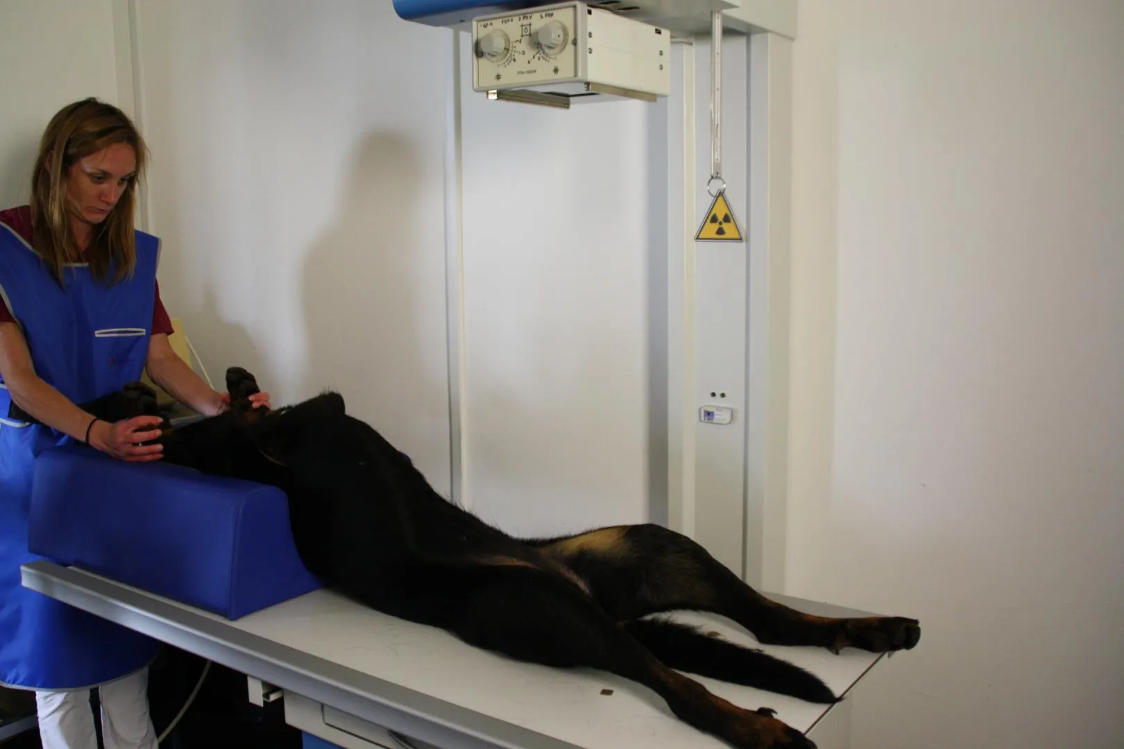
Why does a radiograph taken under anaesthesia allow for a better diagnosis than one taken without?
When the dog is awake for the radiograph, it contracts its muscles — partly because it is stressed (few dogs enjoy being placed on their back with someone pulling their hind legs and another restraining their front legs, all on a table in... a veterinary clinic).
When the dog is anaesthetised (taking care to use muscle-relaxing agents), its muscles relax and no longer participate in holding the femoral heads within the acetabulum: it is then possible to see whether the ligament and articular surfaces are sufficient to hold the heads in the correct position.
When the dog is awake, it is impossible to know whether the hips are truly sound or whether the muscles are compensating for hip defects: when a dog has passive hyperlaxity, its dysplasia cannot be detected radiographically if it is not sedated.
In fact, in these dogs, the only sign of dysplasia is the hyperlaxity, which is masked by contracting muscles. In contrast, a dog with active hyperlaxity will have bone deformities and arthritis, so even without seeing laxity if the dog is awake, bone abnormalities will be visible.
Screening WITH anaesthesia is therefore preferable to screening without anaesthesia for accurately assessing the true state of the dog's hips. To put it simply: a dog with very good hips will obtain roughly the same score with or without anaesthesia. But a dog with average hips has a strong chance of scoring A or B without anaesthesia, whereas in reality its hips are considerably worse and a specialist might grade them C under anaesthesia.
Hip dysplasia is a complex orthopaedic condition that primarily affects medium and large breed dogs. This developmental condition can affect one or both hind limbs, significantly impacting the animal's quality of life.
Genetic predisposition plays a major role in the development of dysplasia, particularly in large breeds. Some breeds are especially affected:
Hip dysplasia is a complex condition that requires a comprehensive approach, from prevention through to treatment. Early detection and appropriate management can optimise the quality of life of affected dogs. Do not hesitate to consult your vet at the first sign of concern — early diagnosis can make a significant difference to the progression of the disease.